A limit in the earlier microscope
The scanning tunnelling microscope, which Gerd Binnig had built with Heinrich Rohrer at IBM Zurich in the early 1980s, worked by measuring a minute electrical current that tunnels between a sharp tip and a surface held a hair’s breadth away, a quantum effect sensitive enough to trace individual atoms. Its central limitation was built into that same mechanism: tunnelling current only flows through conducting or semiconducting materials, so anything insulating, from many ceramics to most biological tissue, simply could not be imaged this way. Binnig, working alongside Christoph Gerber and Calvin Quate, set out to design an instrument that kept the tunnelling microscope’s essential idea, a sharp tip scanning very close to a surface, while replacing the electrical requirement with something that would work regardless of whether the sample conducted electricity.
Feeling instead of tunnelling
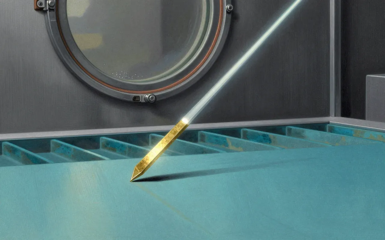
Their answer was to measure force rather than current. A very small, flexible cantilever holds the tip, and as the tip moves across a surface, tiny variations in height cause the cantilever to bend by correspondingly tiny amounts, whether the surface conducts or not. A laser beam reflected off the back of the cantilever onto a position-sensitive detector converts that bending into a signal precise enough to reconstruct the surface’s topography at a resolution reported in fractions of a nanometre, more than a thousand times finer than an optical microscope can manage. Binnig conceived the underlying idea around 1985, and by 1986, working with Gerber and Quate, the team had a functioning instrument, the atomic force microscope, that demonstrated the approach could deliver comparable detail to the tunnelling microscope without its conductivity restriction.
Building it with Gerber and Quate
The core mechanism has held up well: cantilever-based force sensing remains the working principle behind every atomic force microscope built since, refined but not replaced. Three distinct ways of using the tip developed over time, each suited to different samples. Contact mode drags the tip directly across the surface for the most direct measurement, tapping mode oscillates the cantilever so it touches the surface only intermittently, reducing wear and damage, and non-contact mode holds the tip just above the surface entirely, sensing forces without any physical touch at all. That range let researchers choose a mode appropriate to fragile or soft material, something the rigid requirements of the earlier tunnelling technique could not offer.
Reading the bend with a laser
The technique’s resolution and versatility depend heavily on the specific tip, cantilever, and mode chosen, and on the sample itself; delicate biological structures imaged in contact mode can be damaged or distorted by the tip’s direct pressure, which is part of why gentler tapping and non-contact modes were developed in the first place. The instrument also does not replace the tunnelling microscope outright, since for purely conducting samples where extremely fine electronic detail matters, the original tunnelling approach can still offer information the force-based method does not directly provide. The two techniques are better understood as siblings addressing overlapping but not identical problems than as one simply superseding the other.
Three ways to scan
The practical consequence of removing the conductivity requirement was substantial: atomic force microscopy opened nanoscale imaging to insulating materials, semiconductors under processing, polymers, and biological samples including proteins and cell surfaces, none of which the tunnelling microscope could reach. It also let researchers measure mechanical properties directly, stiffness, adhesion, friction, at the nanoscale, rather than only topography, because the cantilever’s response carries information about the forces it encounters, not just the surface’s shape. Commercial instruments became available by 1989, and the technique has since become a standard fixture in materials science, semiconductor research, and molecular biology labs, well beyond the specialised physics settings where the tunnelling microscope first proved the underlying scanning-probe concept.
A tool that reaches biology
This is worth reading selectively rather than as a must, best appreciated alongside the scanning tunnelling microscope story it directly follows from, since much of its interest comes from seeing how quickly a specific technical limitation, no conductivity requirement, generated a specific technical fix, a bending cantilever read by laser. Readers already familiar with the earlier instrument will get the most out of it, watching one invention’s constraint become the design brief for the next. On its own, it is a solid but comparatively narrow account of an engineering solution rather than a conceptual breakthrough, useful background for anyone working with or curious about nanoscale imaging rather than essential reading for a general audience.