9:07in productionCh. 1 · The first sighting/ 9:07 · ceiling 15 min
Life sciences · Medicine
Endoplasmic reticulum
Seeing a structure is not understanding it — and the endoplasmic reticulum took 48 years just to become visible.
The discovery of the endoplasmic reticulum consists of two verified visual milestones: Garnier’s 1897 light microscopic identification and naming of ‘ergastoplasm’, and Porter, Claude & Fullam’s 1945 electron microscopic resolution of its lacy membrane structure. Nothing in the sources describes function, mechanism, biochemistry, or cellular role. The work established visibility — not meaning.
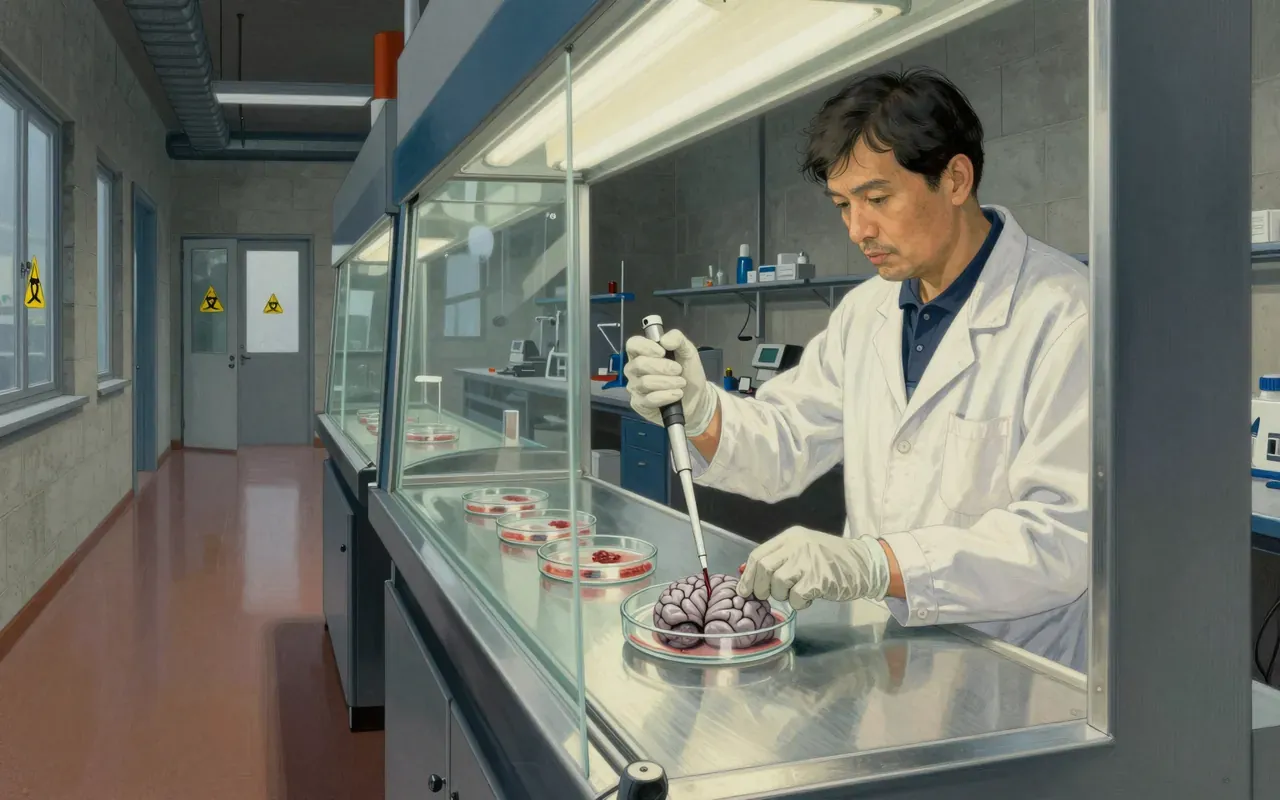
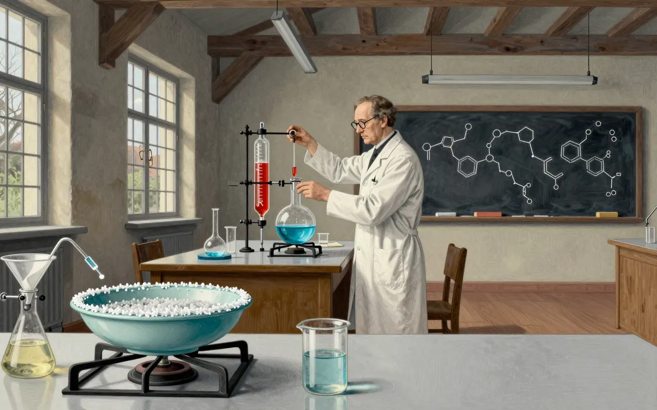
In 1897, Charles Garnier saw a granular network in gland cells under light microscopy and named it ergastoplasm.
2:11
The first resolution
In 1945, electron microscopy revealed the ER’s lacy membrane architecture for the first time.
4:07
Two tools, two truths
Two different instruments — light and electron microscopes — were required to see the same structure at different levels of detail.
5:40
Form without function
Neither observation assigned function — only location, form, and name.
Worth your time?
Yes. Study the whole thing.
4/ 5
What works
establishes a morphological landmark
defines the starting point for organelle biology
exposes the lag between observation and interpretation
What does not
assign function
identify molecular components
link to disease
describe dynamics
Study it if
historians of science
cell biologists reviewing foundations
students learning microscopy limits
Skip it if
clinicians seeking therapeutic targets
biochemists studying protein folding
geneticists mapping organelle inheritance
The written brief1 min read
What the work claims
The work claims only that a distinct intracellular structure was observed and named (1897), then later resolved as a continuous membranous network (1945). It makes no claim about composition, dynamics, or biological activity.
How it was done
Charles Garnier used light microscopy in 1897 to observe a granular network in gland cells. Keith R. Porter, Albert Claude, and Ernest F. Fullam used electron microscopy in 1945 to resolve its lacy membrane structure.
What holds up
Two discrete observational milestones hold: Garnier’s naming of ‘ergastoplasm’ in 1897 via light microscopy, and the 1945 electron micrographic resolution of its lacy membranes.
What does not
Nothing in the sources assigns function, mechanism, biochemical identity, or cellular role to the ER. No claim is made about protein synthesis, calcium storage, lipid metabolism, or disease links.
Why it matters beyond the lab
It matters because every textbook diagram of the eukaryotic cell rests on these two visualisations — but the gap between seeing a structure and knowing what it does remains unbridged by this discovery alone.
Is it worth your time
Yes — it anchors all modern cell biology, but only if you understand that the 1897 and 1945 observations are separate claims about visibility, not function.
Krakatoa didn’t just boom — it turned the atmosphere into a resonant chamber and a reflective shield, with numbers we still use to test climate physics.