A killer that identifies its target from the inside
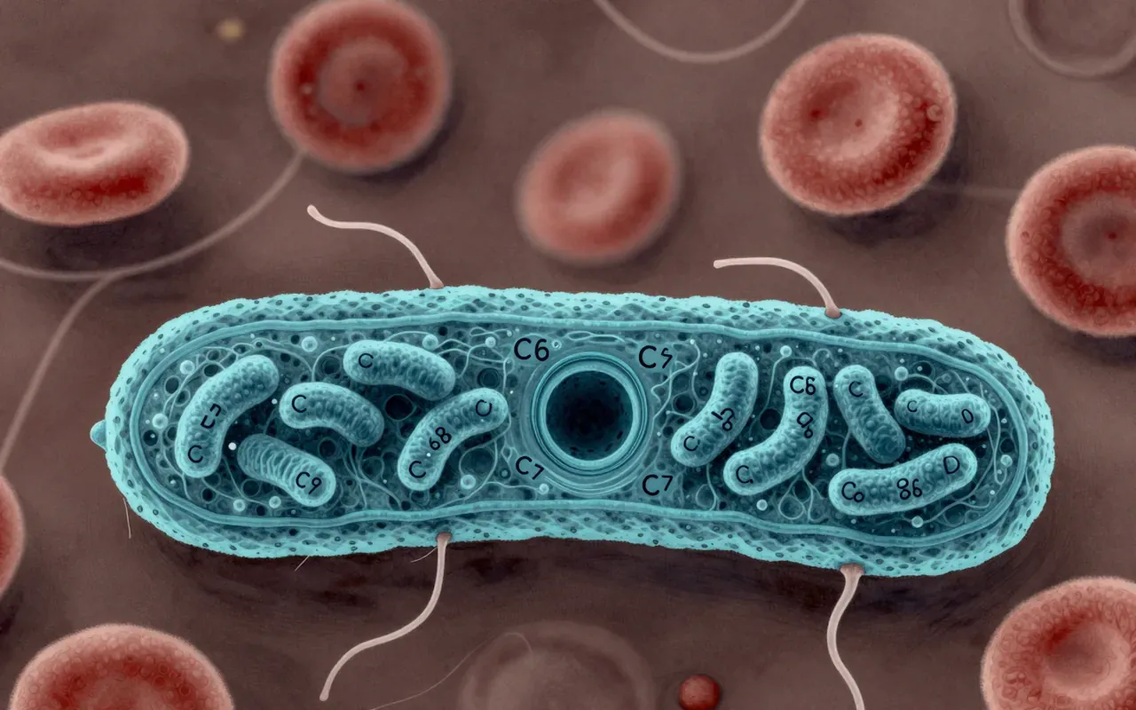
Cytotoxic T cells, also called CD8-positive or killer T cells, are presented here as the immune system’s targeted removal service, eliminating cells that are infected or cancerous rather than attacking pathogens directly in the way antibodies do. What makes their targeting method notable is that it works from the inside out: fragments of proteins made inside a cell are loaded onto MHC class I molecules and displayed on that cell’s surface, and it is this internal signature, not anything visible from outside in a healthy state, that a cytotoxic T cell’s receptor reads. A co-receptor called CD8 stabilises this recognition event. The claim under discussion is that this internal-surveillance system, paired with a controlled killing mechanism rather than a violent one, is what lets the immune system remove compromised cells without simply causing damage wherever it acts.
Two signals before the kill
Before a cytotoxic T cell can kill, it has to be activated, and the material describes this as requiring two separate signals rather than one. The first is the T cell receptor engaging its matching antigen-MHC class I complex, stabilised by CD8. The second is a costimulatory signal, delivered when a molecule on the T cell binds a matching partner on the antigen-presenting cell, typically a dendritic cell. Notably, the material describes dendritic cells needing to interact with both a helper T cell and the cytotoxic T cell for the strongest, most durable activation, with the helper cell effectively licensing the dendritic cell first. The material itself flags this section of the underlying process as one of the harder parts of immunology to explain clearly, which is a useful signal that the two-signal model, while central, is not the whole picture.
Death by instruction, not by force
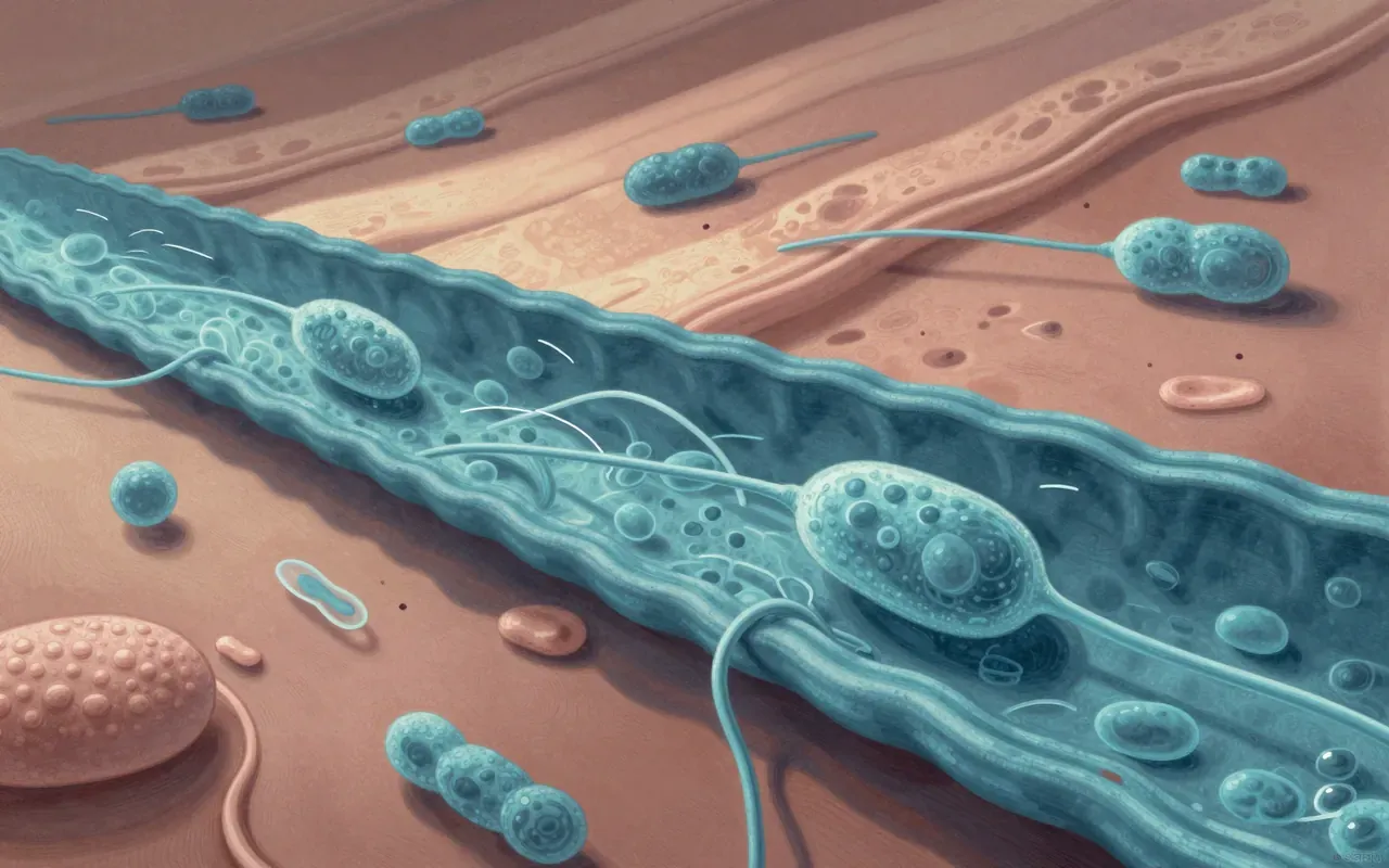
The killing itself happens through two described mechanisms, and neither involves the cytotoxic T cell physically tearing its target apart. The primary route delivers perforin, a protein that creates an opening in the target cell’s membrane, and granzymes, enzymes that pass through that opening into the target’s cytoplasm and activate a cascade of proteases called caspases, setting off apoptosis within a matter of hours. A second route uses a surface protein called Fas ligand, which binds a receptor on the target cell and assembles a death-inducing complex that activates the same caspase cascade by a different entry point. Cytotoxic T cells are described as resistant to their own granzymes, owing to specific features of their membrane, which is what lets them deliver this signal repeatedly without dying alongside their targets.
A word coined for falling leaves
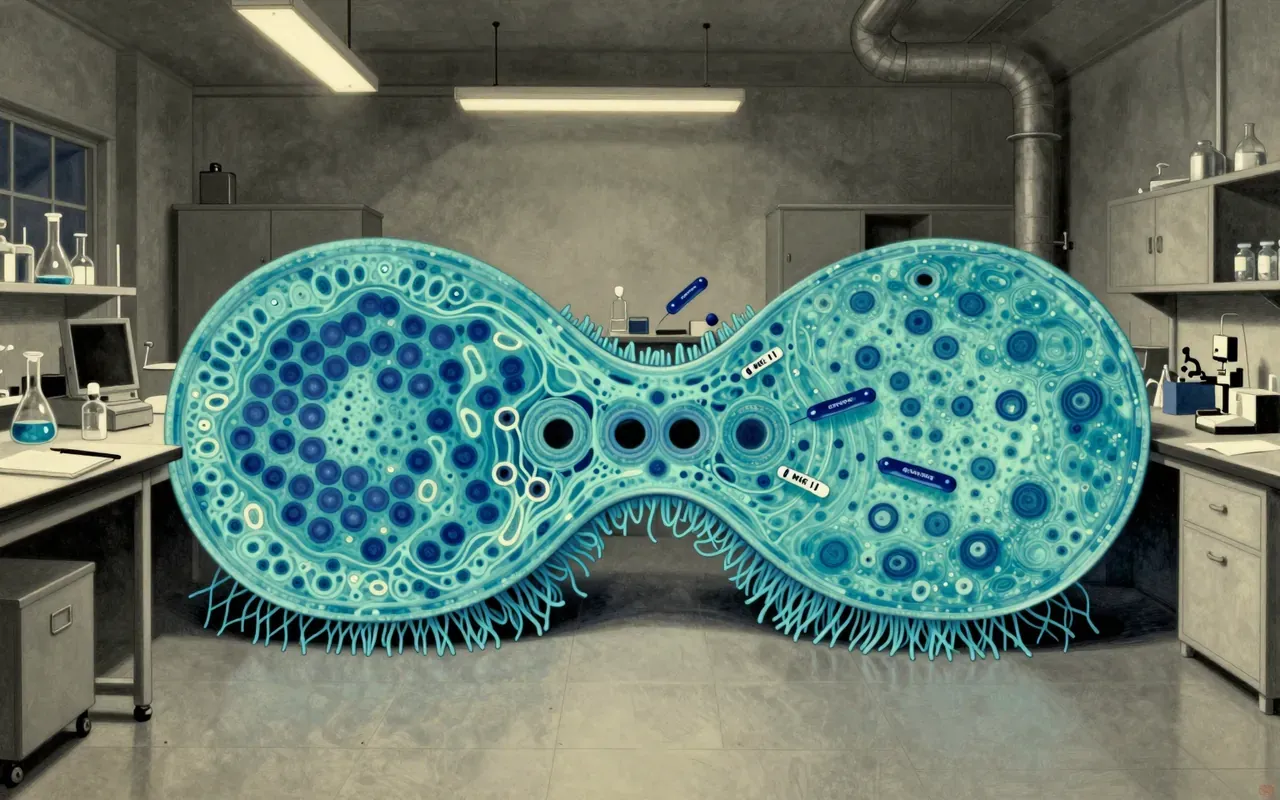
Apoptosis, the death programme cytotoxic T cells exploit, was formally distinguished from ordinary traumatic cell death in 1972, when John Kerr, Andrew Wyllie and Alastair Currie published a paper describing a form of cell death that was orderly, contained, and clearly different from the messy rupture of necrosis. The name itself came from a Greek professor’s suggestion, drawing on a term used since antiquity for leaves falling from a tree, chosen to capture how apoptotic cells shrink and break into contained fragments that neighbouring cells clean up, rather than spilling their contents and provoking inflammation. Decades later, work identifying the specific genes controlling this process in the roundworm Caenorhabditis elegans earned Sydney Brenner, H. Robert Horvitz and John Sulston the Nobel Prize in Physiology or Medicine in 2002.
The same pathway, everyday use
The material is emphatic that apoptosis is not primarily an immune weapon at all, but a routine developmental tool that cytotoxic T cells have essentially borrowed. The same caspase cascade that clears a virus-infected cell also separates individual fingers and toes during embryonic development, removing the tissue between them on a genetically programmed schedule, and drives the dramatic loss of a tadpole’s tail and gills during metamorphosis. This framing matters for understanding what a cytotoxic T cell is actually doing when it kills: rather than deploying a novel destructive process, it is triggering machinery the target cell already carries and would, in different circumstances, use on itself. That shared machinery is also why the same pathway shows up repeatedly across unrelated areas of biology and disease.
When the system fails in either direction
Because too little or too much of this process both cause disease, the balance point is where the material’s stakes become concrete. Cancer cells frequently survive by disabling parts of the apoptotic pathway, keeping a gene called BCL2 active when it should allow death to proceed, which is presented as a central mechanism behind unchecked tumour growth. In the opposite direction, HIV drives excessive apoptosis specifically among CD4 T helper cells, contributing directly to the immune collapse seen in AIDS, and excessive apoptosis is also implicated in neurodegenerative disease. This is a genuinely rewarding read for anyone who wants the mechanics behind two terms, killer T cell and apoptosis, that circulate constantly in medical reporting without ever being explained together, and it earns the hour by showing they are one connected story rather than two separate ones.