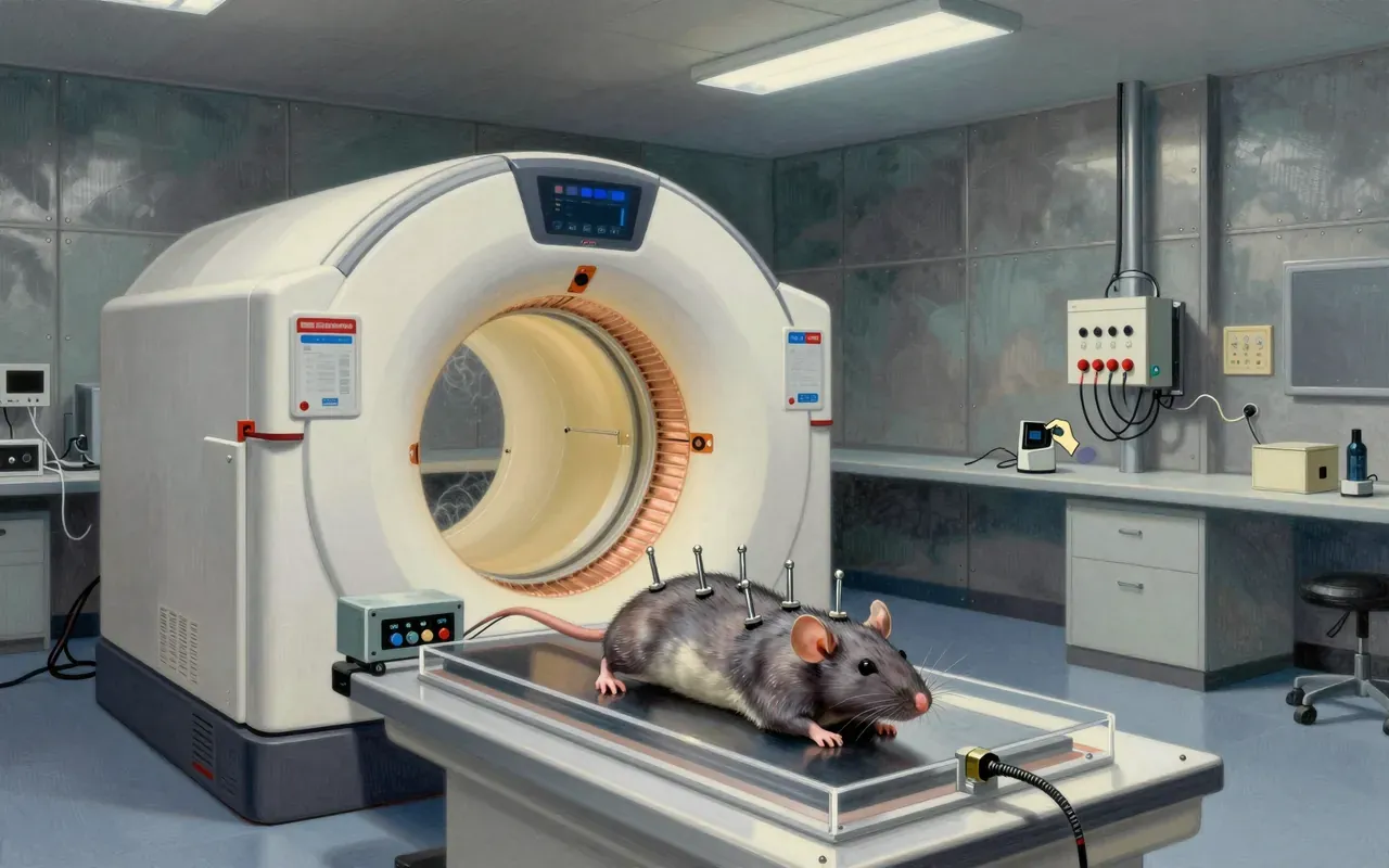
fMRI doesn’t read minds — it maps blood’s magnetic response to oxygen, and Ogawa proved it in rats.
Ogawa’s 1990 work established the BOLD effect — a measurable, oxygenation-dependent MRI signal shift in rodent brain tissue — verified with blood samples and EEG correlation. It is the physical foundation of human fMRI, but maps vascular response, not neural activity directly.
Ogawa discovered the BOLD effect — the first demonstration that functional brain imaging depends on blood oxygenation status.
2:40
The apparatus
They used 7.0 T MRI and gradient-echo imaging to detect oxygenation-dependent signal changes in rodents.
3:52
The experiment
By altering inhaled oxygen and recording EEG, they tied MRI signal shifts to functional brain activity — not just anatomy.
5:27
The verification
They confirmed the effect ex vivo with blood-filled test tubes — proving oxygenation alone alters MRI signal.
6:51
The extension
This principle became the basis for noninvasive functional mapping of the human brain — extending structural MRI into physiology.
9:08
The limit
The resulting map reflects where neurons respond electrochemically — but only indirectly, via blood flow.
Worth your time?
Yes. Study the whole thing.
4.5/ 5
What works
establishes BOLD as physical basis for fMRI
enables noninvasive functional mapping in humans
provides reproducible, ex vivo–verified mechanism
What does not
measures neuronal firing directly
quantifies temporal dynamics
applies to humans in the 1990 study
Study it if
neuroscientists
radiologists
cognitive scientists
Skip it if
clinicians seeking diagnostic biomarkers
philosophers of mind seeking neural correlates of consciousness
The written brief1 min read
What the work claims
That changes in blood oxygenation alter MRI signal properties, enabling a noninvasive map of functional brain activity — specifically, which neurons respond electrochemically to mental processes — via blood-flow correlates.
How it was done
Ogawa and colleagues used 7.0 T MRI to scan rodents while manipulating inhaled oxygen levels. They verified the signal change using test tubes of oxygenated and deoxygenated blood. They identified gradient-echo/T2*-weighted imaging as optimal. They correlated MRI signal changes with EEG-recorded brain activity during gas manipulation.
What holds up
The BOLD effect — MRI signal changes tied to blood oxygenation — was reproducibly observed in rodents, verified ex vivo with blood samples, linked to physiological perturbation (gas manipulation), and correlated with electrophysiological activity (EEG). Gradient-echo imaging was shown optimal for detecting it.
What does not
The work does not show that fMRI measures neuronal firing directly. It does not quantify timing, spatial resolution, or causal links between oxygenation changes and specific mental processes. It does not extend to human subjects in the 1990 study.
Why it matters beyond the lab
It enabled noninvasive functional brain mapping in humans, extending structural MRI into physiology. But it maps haemodynamic response, not cognition — a crucial distinction for interpreting studies on decision-making, emotion, or disease.
Is it worth your time
Yes — it established the physical basis for noninvasive functional brain mapping in humans, but only via the BOLD effect’s vascular proxy, not direct neuronal measurement.