A microscope from an AI pioneer
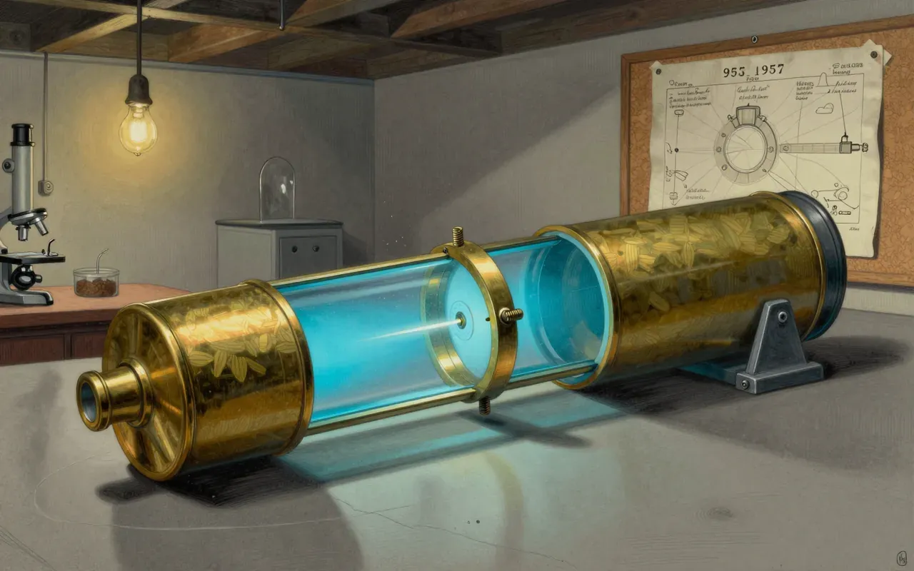
In 1955, Marvin Minsky, then a young mathematician at the very start of what would become a long career in artificial intelligence, built the first confocal scanning microscope, later filing a patent describing the principle in 1957. No images from his original instrument survive, and Minsky never published a scientific paper describing it, an unusual origin for a technology that would eventually become a standard tool across biology and medicine, invented as something of a side project by someone whose scientific reputation would end up resting almost entirely elsewhere, in a field that had nothing to do with optics or imaging at all.
A pinhole that blocks the blur
The core idea is a pinhole aperture placed in front of the detector, positioned at a plane optically matched to the microscope’s focal plane, so that light coming from anywhere except the exact point currently in focus is physically blocked before it can reach the detector, rather than being recorded and blurring the image the way it does in an ordinary microscope. To build a full picture rather than a single point, the instrument scans a point of illumination across the sample, typically using one or more oscillating mirrors to sweep the beam, reconstructing a two- or three-dimensional image point by point rather than capturing the whole field of view in one exposure.
Point by point, layer by layer
Because only light produced very close to the exact plane of focus can reach the detector at any given moment, confocal images achieve markedly better optical resolution than conventional widefield microscopes, especially in the depth direction, letting researchers capture a genuinely sharp image of one thin layer of a sample without light scattered from other depths smearing it. Scanning through a series of focal depths and combining the results, a technique called optical sectioning, produces a three-dimensional reconstruction of a specimen’s internal structure without physically cutting it into slices, a capability that remains the central reason the method is used at all.
Sharper than looking at everything at once
Minsky’s original design went essentially unused for years, largely because the light sources available in the 1950s were not strong enough to make point-by-point scanning practical at any useful speed. The technology only became genuinely usable once other researchers built on the underlying principle with better light sources. M. David Egger and Paul Davidovits described the first confocal laser scanning microscope in papers published in 1969 and 1971, using a 5-milliwatt helium-neon laser to image nerve tissue, and Mojmír Petráň developed the first commercially available confocal instrument, the Tandem Scanning Microscope, filing a Czechoslovak patent for the design in 1966.
Decades on a shelf, waiting for a laser
Further refinements followed steadily rather than all at once. Colin Sheppard and colleagues at Oxford described confocal systems using laser illumination and stage scanning between 1978 and 1980, and William Bradshaw Amos and John Graham White built the first confocal beam-scanning microscope at Cambridge in the mid-1980s, roughly three decades after Minsky’s original patent, before laser technology and detector sensitivity had matured enough to make the method practical for everyday laboratory use. Once it was, confocal laser scanning microscopy spread quickly into cell biology, genetics, microbiology and developmental biology, and clinically into evaluating eye diseases and imaging the cells lining the cornea.
From eye clinics to cell biology labs
This is worth an hour for the unusually long and quiet gap between the idea and its adoption, an instrument that sat essentially unused for roughly three decades not because the underlying principle was wrong but because the supporting technology, sufficiently strong and controllable light sources, had not yet caught up to it. Minsky, remembered almost entirely for his contributions to artificial intelligence and a co-founder of what became MIT’s Computer Science and Artificial Intelligence Laboratory, never returned to the microscope himself, leaving its eventual adoption in biology and medicine to researchers working decades after his original patent had already faded into obscurity.