The crystallisation problem
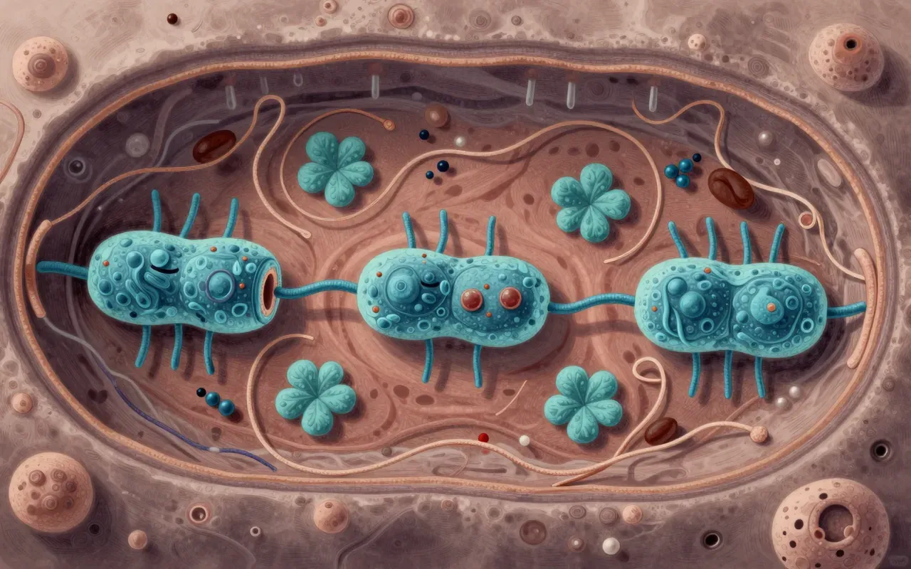
For decades, the standard way to see a protein’s atomic structure was X-ray crystallography, which requires coaxing the molecule into a regular, repeating crystal lattice before firing X-rays through it. That step works well for some proteins and is nearly impossible for others, particularly the membrane proteins that sit half-embedded in a cell’s outer wall and tend not to hold a stable crystal shape at all. Richard Henderson, working at Cambridge’s Laboratory of Molecular Biology from the early 1970s, pursued an alternative: using electron microscopy directly on the protein instead, without requiring a crystal. His early collaboration with Nigel Unwin on bacteriorhodopsin, published in 1975, produced a low-resolution model showing the protein arranged as seven helices crossing the membrane, evidence that membrane proteins had definite, mappable structures after all.
Freezing without ice crystals
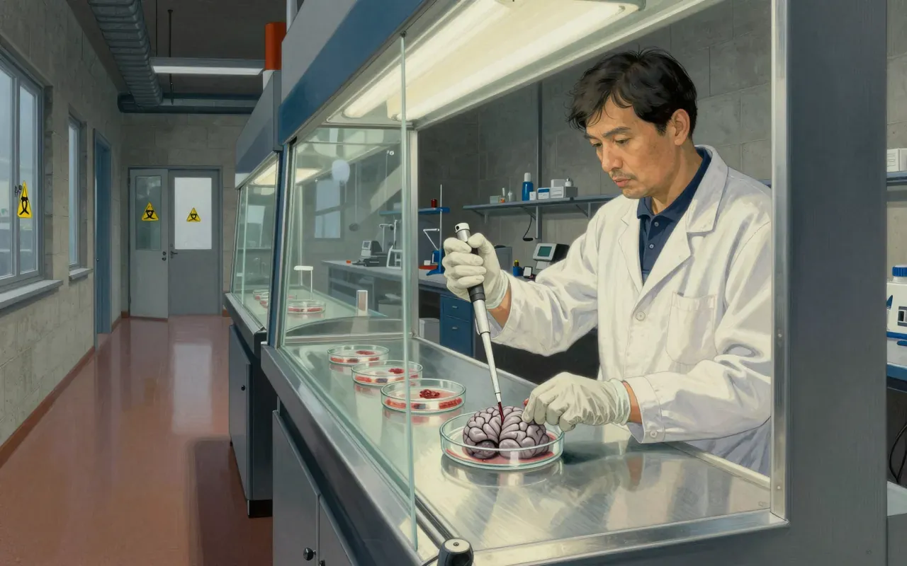
The core technical problem was that ordinary freezing turns water into crystalline ice, and ice crystals distort or destroy the delicate arrangement of a biological molecule before it can be imaged. The solution, developed and refined over subsequent years by Henderson and others, was to freeze samples fast enough that the water forms vitreous ice instead, a disordered, glass-like state that traps the molecule roughly as it existed in solution rather than crushing it into a crystal lattice. Henderson pushed his own bacteriorhodopsin work toward ever finer detail, and by 1990 he had produced an atomic-resolution model of the protein using electron crystallography, only the second atomic-level structure ever obtained for a membrane protein by any method, a result that argued the electron microscopy route could in principle match X-ray crystallography’s precision.
A membrane protein in 1975
The method Henderson championed held up, eventually, as a genuine and now widely used alternative for exactly the cases X-ray crystallography struggles with. The turning point came around 2012 and 2013, when direct electron detectors, cameras that record electrons hitting them far more efficiently than earlier film or indirect sensors, arrived alongside improved computational algorithms for combining thousands of noisy individual images into one sharp structure. Together these advances produced what researchers have called a resolution revolution, and cryo-EM structures reaching near-atomic detail, and in some cases resolutions as fine as roughly 1.2 Ångströms by 2020, became achievable for viruses, ribosomes, ion channels, and large enzyme complexes that had previously resisted structural study by any technique.
Atomic resolution by 1990
What the method still does not fully replace is X-ray crystallography’s usefulness for very small, well-behaved molecules that crystallise easily and can be resolved to extremely fine detail that way with comparatively modest equipment. Cryo-EM’s advantage grows with the size and structural awkwardness of the target, and it took a long stretch of methodological work, spanning Henderson’s own career from the 1970s through the 2010s, before the computational and hardware pieces caught up with the underlying idea. The gap between having a working concept and having a practical, widely deployable tool was measured in decades, not years, which is easy to lose sight of when the technique now appears as a routine part of structural biology papers.
The resolution revolution
The wider consequence is that structures once considered essentially unreachable, including many membrane proteins that are the targets of a large share of modern drugs, became visible in detail without needing to solve the separate, often intractable problem of growing them into crystals. That has fed directly into drug design and into basic understanding of how large molecular machines, ribosomes among them, actually assemble and function. The 2017 Nobel Prize in Chemistry, shared by Henderson with Jacques Dubochet, who worked out the vitrification freezing method, and Joachim Frank, who developed key image-processing approaches, recognised that the achievement was distributed across several complementary contributions rather than belonging to any one person’s single breakthrough.
A shared Nobel Prize
This is worth understanding because it is an unusually honest example of how a scientific method actually matures: not through one triumphant experiment but through a long sequence of partial results, from a rough 1975 helix map to a 1990 atomic structure to a 2012 hardware leap, each step necessary and none sufficient on its own. Readers drawn to dramatic single discoveries may find the pace slower than they expect, since the interesting part is the accumulation rather than any one moment. But for anyone who wants to understand why cryo-EM structures now appear so routinely in biology and medicine, and why it took until the 2010s rather than the 1970s for the promise Henderson saw early to actually pay off, it is time well spent.