Two routes to the same idea
Computed tomography is the claim that a full cross-sectional image of the inside of the body can be reconstructed from a series of X-ray measurements taken from many different angles around it, without needing to see through the body in a single pass the way a conventional X-ray does. Godfrey Hounsfield, an electrical engineer at EMI, arrived at the idea independently while thinking about how to determine the contents of a box from readings taken all around it, then built a working scanner to test it. Allan Cormack, a physicist, had derived much of the underlying mathematics for reconstructing an image from angled measurements years before Hounsfield’s engineering work, arriving at a closely related idea from theory rather than from building a device.
The first scan
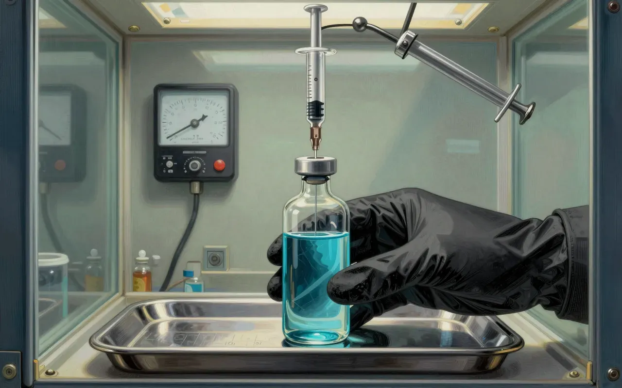
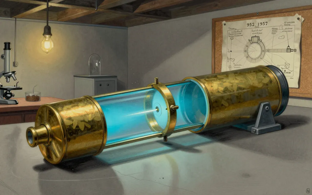
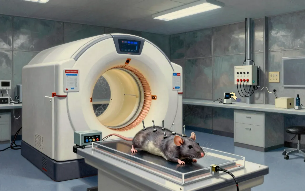
Hounsfield tested his approach first on preserved brain tissue, then on fresh specimens, refining the scanning and reconstruction process before attempting it on a person. The first successful scan of a patient took place on 1 October 1971 at Atkinson Morley Hospital in Wimbledon, imaging a patient with a suspected cerebral cyst. The scanner worked by rotating an X-ray source and detector around the head, taking many individual absorption readings at different angles, then using tomographic reconstruction — a computational method for working backwards from a set of angled projections to the arrangement of tissue that produced them — to assemble those readings into cross-sectional slices. Hounsfield went on to build a whole-body version of the scanner, extending the same principle beyond the head, by 1975.
One principle, decades of use
The reconstruction principle itself has held completely: every CT scanner built since works on the same basis of combining many angled X-ray measurements into cross-sectional images, refined in speed and resolution but not changed in kind. Cormack’s mathematics, developed independently of Hounsfield’s engineering, held up as the theoretical grounding for why the reconstruction method works at all, which is why the two men shared the 1979 Nobel Prize in Physiology or Medicine rather than the prize going to the engineering achievement alone. The clinical usefulness of the resulting images has also held up across an enormous range of applications the material lists — detecting stroke, tumours, bone trauma, pulmonary embolism, coronary artery disease and abdominal conditions — making CT one of the most broadly used diagnostic tools to emerge from twentieth-century physics and engineering combined.
The dose that comes with the image
What the material is careful to record rather than gloss over is the radiation cost of the technique: a routine abdominal CT delivers a dose the source compares to roughly three years of background radiation, which is not a trivial exposure and is the tradeoff against the images’ diagnostic value. The scale of use compounds that concern rather than dissolving it — an estimated 72 million CT scans were performed in the United States in 2007, rising to over 80 million by 2015, meaning cumulative population exposure from the technology has grown substantially even as individual scans have not necessarily become riskier. Nothing here suggests the reconstruction principle itself is in doubt; the caution is squarely about dose and frequency of use, not about whether the images are trustworthy.
From the head to the whole body
CT reconstruction is now one of the standard tools of diagnostic medicine, supplementing ordinary X-ray and ultrasound in situations where a cross-sectional view changes what a clinician can see — internal bleeding, tumour location, complex fractures, or blockages that a flat image cannot resolve. Beyond clinical medicine, the same mathematics has migrated into non-medical imaging of objects, using X-rays to see inside manufactured parts or archaeological materials without cutting them open. The underlying story is also a useful example of two independent routes reaching the same result: an engineer solving a practical problem by building and testing, and a physicist solving the equivalent problem on paper, converging on one working method that neither could have delivered to the same effect alone.
Theory and engineering meeting in the middle
Worth understanding for what it shows about how theory and engineering can arrive at the same destination from opposite directions — Cormack’s mathematics sitting unapplied for years until Hounsfield’s scanner gave it a working machine to describe, and Hounsfield’s machine needing Cormack’s mathematics to be more than an empirical curiosity. It is a clean, well-documented case of an invention with a clear before-and-after: medical imaging that could only look through the body in flat projection, and then, from 1971, imaging that could see inside it in cross-section. Anyone wanting a rundown of exactly how modern CT hardware differs from Hounsfield’s 1971 prototype will need to look elsewhere, since the source material stays with the original invention and its clinical consequences rather than tracing the technology’s later engineering refinements.