A cell at a time
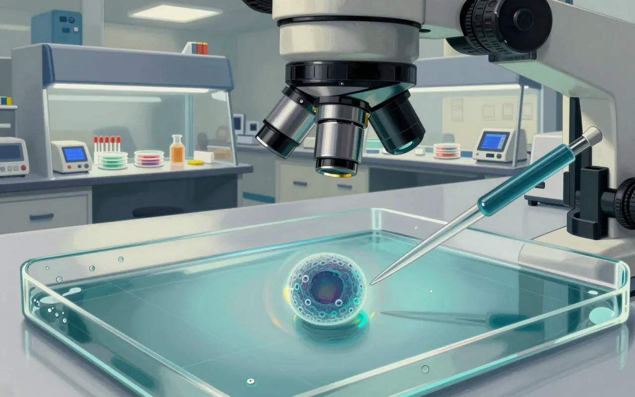
Flow cytometry works by suspending cells or other particles in fluid and passing them, one at a time, through a narrow stream where they cross the path of a laser beam. As each individual cell passes through that beam, the instrument records how the light scatters off it and how much fluorescent light it gives off, then processes those optical signals with a computer to build up a detailed picture of thousands of individual cells’ physical and chemical characteristics, all within seconds rather than the hours a manual equivalent would require.
Measuring resistance, not light
The technology’s roots go back further than laser-based fluorescence, to Wallace Coulter’s 1953 patent, US Patent 2,656,508, describing a different detection principle, since called the Coulter principle, which measured cells by the tiny change in electrical resistance each one caused while passing through a small aperture. This was a purely electrical rather than optical way of counting and sizing individual particles in a fluid stream, and it predates any of the fluorescence-based features that define modern flow cytometers by well over a decade, working purely on electronics rather than optics of any kind.
An ink-jet printer and a cell sorter
The idea of not just counting cells but physically sorting them by their measured characteristics came from Mack Fulwyler, who in 1965 built the first prototype cell sorter at Los Alamos National Laboratory by combining a Coulter volume sensor with an ink-jet printer mechanism, publishing the result in the journal Science. His design let individual cells, once measured, be diverted and physically separated from one another based on that measurement, rather than simply being counted and discarded, turning a purely analytical instrument into a genuine separation tool for the first time.
Fluorescence enters the picture
Fluorescence detection, now the technique’s defining feature, arrived a few years later. Wolfgang Göhde at the University of Münster built the first fluorescence-based instrument, the ICP 11, in 1968, filing a patent for it that December, with the German company Partec bringing a commercial version to market around 1968 and 1969. Around the same period, Len Herzenberg at Stanford developed the fluorescence-activated cell sorter, combining fluorescence detection with Fulwyler’s sorting concept into a design that shaped most modern instruments, work later recognised with the 2006 Kyoto Prize. The field only settled on the name flow cytometry itself in 1976, at a conference in Pensacola, Florida, eight years after Göhde’s instrument had already appeared.
Thousands of cells a second, sorted by charge
Modern flow cytometers keep individual cells centred in a narrow stream using an outer layer of sheath fluid, a technique called hydrodynamic focusing, sometimes assisted by acoustic waves above 2 megahertz to pre-align particles before they reach the laser. Commercial instruments can now carry as many as ten separate lasers and thirty fluorescence detectors, splitting scattered and emitted light by wavelength through filters, dichroic mirrors and diffraction gratings onto photomultiplier tubes or avalanche photodiodes. When sorting rather than simply measuring, the stream breaks into droplets at a vibrating nozzle, typically holding one cell or none each, and an electrical charging ring assigns each droplet a charge based on what was just measured, letting an electric field steer it into the correct collection container.
From cancer wards to marine biology
The result is used across an unusually wide range of fields, from immunology, virology and plant biology to marine biology, and clinically for diagnosing blood cancers, monitoring the effects of chemotherapy, prenatal diagnosis, transplant medicine and sperm sorting, typically processing many thousands of individual cells per second in real time. This is worth an hour for how directly the instrument’s two founding ideas, Coulter’s electrical detection of a passing particle and Fulwyler’s ink-jet-based physical sorting of one, both borrowed from entirely unrelated industrial technology, were combined and refined over decades into a single machine now central to cancer diagnosis and basic cell biology alike.